Petri Dish Bacteria Identification Chart


On a nonporous surface ie Petri dish or glass plate wet a strip of filter paper with a few drops of Kovacs oxidase reagent.
Petri dish bacteria identification chart. Mar 6 2018 - Bacteria grow on solid media as colonies. To identify bacteria and fungi from their environment. 4 5 Various Gram-negative bacterial colonies growing on MacConkeys agar.
For long term storage one loop of bacteria was mixed in. The Gram stain test allows scientists to classify bacterial shapes by analyzing their cell walls. Flow chart for identification and characterization of a N.
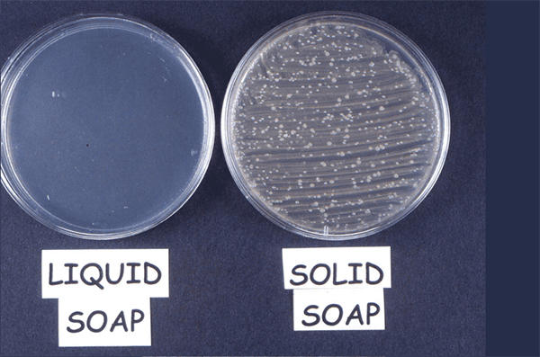
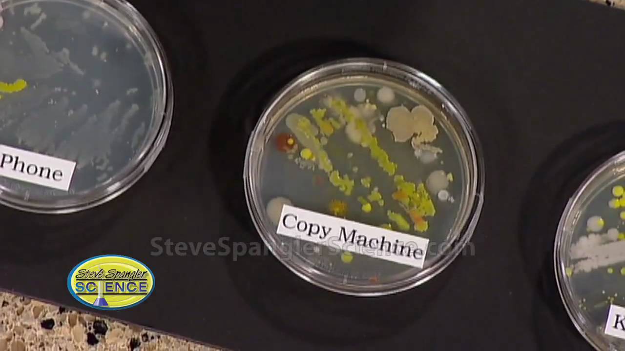
After we cultured our petri dishes with swabs from several household items we set the dishes under a desk lamp to keep the temperature around 95 degrees F 35 degrees C. We should examine the bacteria colonies in the closed petri dish for safety. Touch plates growing bacteria from the fingertips.
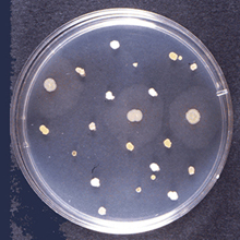
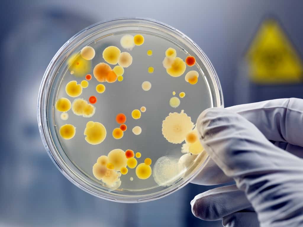
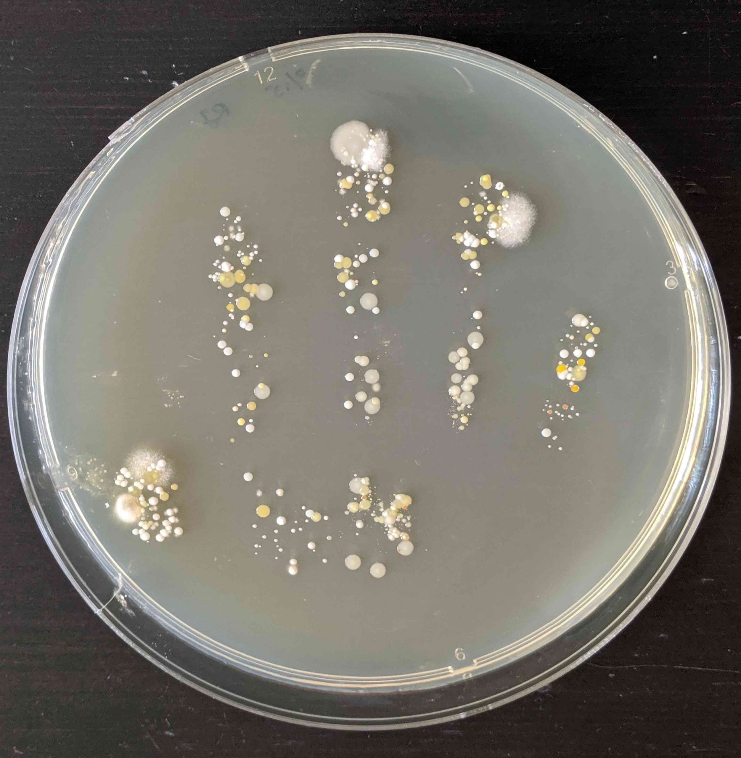
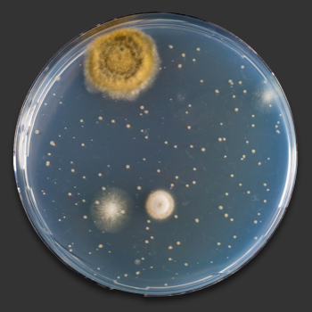
Colony morphology is a method that scientists use to describe the characteristics of an individual colony of bacteria growing on agar in a Petri dish. Coccobacilli elongated coccal form. Moraxella catarrhalis Gram stain.
Cocci sounds like cox-eye spherical spirillum pl. Klebsiella pneumoniae and Saureus Gram stain. Remember that a laboratory.
Microscopy and automated system for rapid identification of bacteria BioLog identification system. There will likely be many different types of bacteria in a clinical sample so some of the bacterial colonies that grow will need to be isolated and tested to determine which type of bacteria is. Colony morphology is a method that scientists use to describe the characteristics of an individual colony of bacteria growing on agar in a Petri dish.
We may not see them but microbes are all around. The dish is partially filled with warm liquid containing agar and a mixture of specific ingredients that may include nutrients blood salts carbohydrates dyes indicators amino acids or antibiotics. As well as other harmful pathogens that are not mold such as bacteria that were not detected by your culture plate.
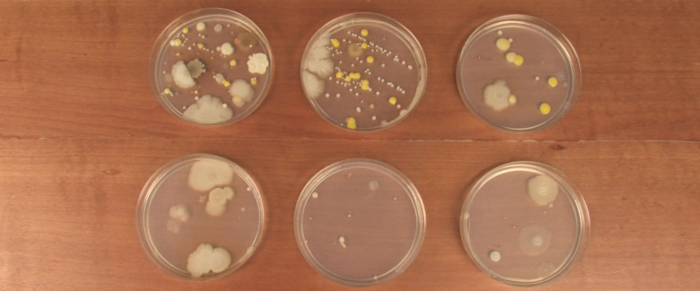
There are many ways in which you can identify the bacteria. The petri dishes were incubated for 5 days at 281 C in an incubator with a 12-h light cycle. The bacteria thus isolated needs to be further identified to genus and.
Bacilli rod-shaped coccus pl. Armed with cotton swabs and Petri dishes full of nutient agar. It is virtually impossible to completely remove mold spores from an indoor environment but.
EVVIVA SCIENCES MOLD IDENTIFICATION GUIDE 01. The text is intended as a reference guide to help building owners operate and maintain their home effectively. One of the most common ways to identify the 3 shapes of bacteria is through the Gram stain test.
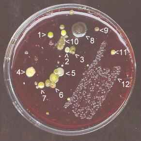
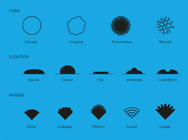
Use a disposable plastic loop a platinum inoculating. By Lifestyle February 25 2021 1045 am. Measuring the diameter of the colony Shape.
No acid production in glucose and maltose andor acid production in sucrose or. Spirilla spiral Some bacteria have more unusual shapes. Understanding the Different Types of Bacteria.
Photos from the SPO Bacterial Media Culture Laboratory Exercise. MODULE Bacterial Identification Tests Microbiology 122 Notes 11 BACTERIAL IDENTIFICATION TESTS 111 INTRODUCTION In the previous chapter we have discussed various methods of isolation of bacteria. Inside the Petri Dish.
Petri-dish and 20 ml of MRS agar was then. Petri dishes are often used to make agar plates for microbiology studies. If the temperature is too hot over 100 degrees F 37 degrees C some types of.
Students should examine cultures in containers which have been taped and closed. Neisseria gonorrhoeae on New York City Agar. It is useful for bacteria identification.
Identification by Shape Some of the first steps in identifying bacteria are to examine according to shape. Colony morphology A swab from a bin spread directly onto nutrient. Moraxella catarrhalis on blood agar.
Observing bacteria in a Petri dish. Normal flora from the arm. The culture was kept in MRS agar slant and stored at 4 C.
If the temperature is too low bacteria will still grow but the process will be very slow. First and foremost it can be done visually by looking at the colonies ie the cultural characteristics and noting the following Size. A clinical sample of bacteria is obtained from the patient and then platedstreaked onto a Petri dish of bacterial growth medium and incubated for growth.
The identification was performed according to Bergeys manual of determinative of bacteriology 14. Let the filter paper strip air dry before use. Additional Sources and Resources.
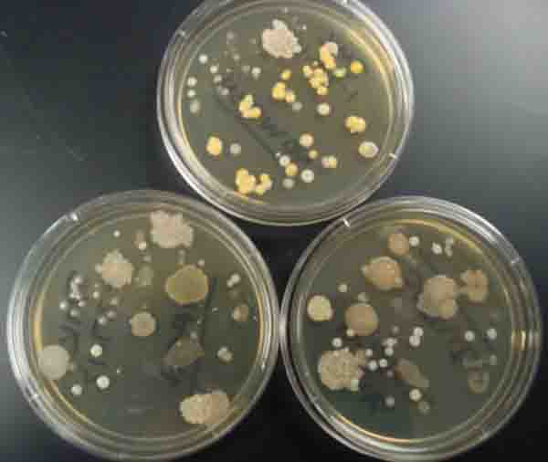
This fact is revealed to microbiology students who are tasked with a classic project. Using a sterile inoculating loop or wooden. Bacteria colonies differ in their shape size color and margin.
A Petri dish with bacterial colonies on an agar-based growth medium. A colony is defined as a visible mass of microorganisms all originating from a single mother cell therefore a colony constitutes a clone of bacteria all. The Good Cause Guide.
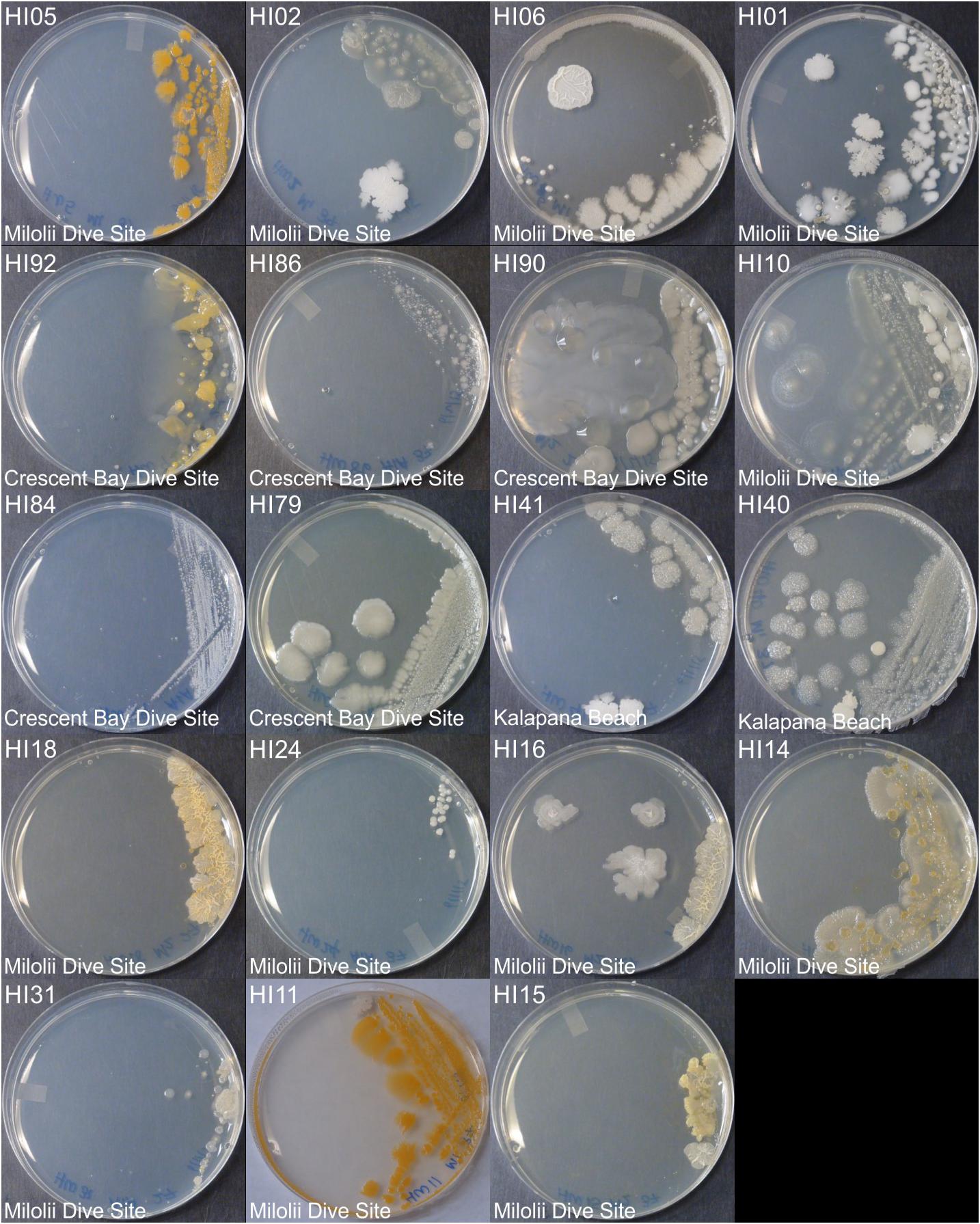
The images below show the appearance of many common household molds on petri dishes. Obtained using an arm plate. Observing bacteria colonies in a Petri dish.
Identification of Bacteria. It can be used to help to identify them. Each grain sample 200 grains treatment 1 was examined to identify fungi up to the species level.
Grains with and without surface sterilization were transferred separately to pre-sterilized petri dish humid chambers under aseptic conditions. Compare the mold growth from your kit with the images below to help identify the mold on your petri plates. Keep the petri dish cover available.